What would be required if we were seriously to contemplate the replacement of the chest x ray with CT scanning in the acute setting?
 The medical registrar was on the phone for the third time that morning. “Another CTPA?” I asked, aiming for a tone of polite surprise, but probably failing. “Haven’t you heard?” she said, “It’s the new chest x ray.”
CT pulmonary angiography (CTPA) was introduced into clinical practice about 20 years ago. An accurate test for a common condition—pulmonary embolism—which can present acutely, is known to be hard to diagnose, and is sometimes fatal, has an obvious appeal. And unlike the traditional alternative—the perfusion lung scan—it carries the advantage of being able to suggest or exclude a range of other possible diagnoses. It has proved to be a very popular test, to the point at which concerns have been raised that its use may have resulted in potentially harmful overdiagnosis.
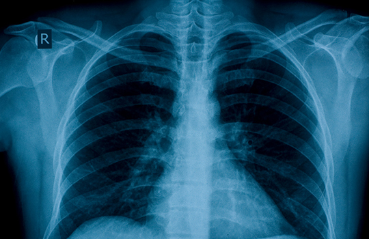
The chest x ray has been around for a long time and familiarity can perhaps blind us to its deficiencies, of which the inability to diagnose pulmonary embolism is only one. For example, about a quarter of lung cancers and half of all rib fractures are not detected on chest x ray. It makes no contribution to the diagnosis of asthma, coronary heart disease, or a range of other cardiorespiratory conditions until severe complications have developed. It provides very little functional information and, to put it bluntly, many people die following a normal chest x ray. And yet it retains a central role in the assessment of patients with acute medical problems, almost a rite of passage in the process of admission to hospital.
Over recent decades, the use of plain radiographs in many other clinical circumstances has come under increasing scrutiny. The limitations of the abdominal x ray in patients with acute abdominal pain are now widely accepted as the greater use of ultrasound, and particularly CT scanning, has made its deficiencies glaringly apparent. X rays of the spine for trauma or back pain are now only indicated in specific circumstances according to the most recent guidance, replaced when necessary by CT or MRI in selected patients when imaging is essential to managing a patient’s treatment. For how much longer will we continue to put the chest x ray at the heart of acute medical diagnosis?
It may be worth thinking about what would be required if we were seriously to contemplate the replacement of the chest x ray with CT scanning in the acute setting. To start with, we would need a new generation of low cost, accessible, low dose CT scanners, installed in every emergency room to produce the necessary increase in scanning capacity. The traditional brake on the use of CT scanning has been appropriate concern over the implications of the higher radiation dose involved. Technological developments in dose reduction—to the point at which CT images can be acquired with doses approaching those of a chest x ray—are challenging the relevance of this.
We would need staff trained in the use of these machines, and many more staff trained to interpret the images. Existing programmes for teaching chest x ray interpretation to a wide range of healthcare practitioners would need to be augmented with CT teaching.
Less obviously, we would need an increased understanding and tolerance of the inevitable incidental findings which would proliferate—an appreciation, for example, that a high proportion of adults admitted with acute medical conditions will have at least one small pulmonary nodule and that enlarged mediastinal lymph nodes are found in most acute conditions and only rarely indicate malignancy. A test which inevitably generates serial follow-up imaging would be of limited value.
Perhaps most importantly, we would require the establishment of an evidence base to show that CT does genuinely out-perform the chest x ray in improving outcomes for unselected acute patients without causing additional harm. How well for example does CT scanning perform in answering the question posed by acute physicians everywhere—is this patient’s breathlessness caused by infection, pulmonary oedema, both or neither? Evidence at the moment is in short supply.
Is it conceivable that we could ever kick the chest x ray habit and employ more sophisticated imaging for those patients whose management really depends on the outcome? I don’t know, but looking at the list above, I suspect that the new chest x ray may still be a little way off.
Giles Maskell is a radiologist in Truro. He is past president of the Royal College of Radiologists.
Competing interests: None declared.