An article recently published in Veterinary Record Case Reports describes the case of a 10-year-old, male, intact Border collie presented with a 12-month history of progressive paraparesis and lumbar discomfort and a 5-year-old male neutered, domestic short-hair cat presented with a one-week rapid progressive history of unusual stance and gait. Both pets adopted a ‘praying position’-like posture and a gait characterised by raising the pelvis while keeping the elbows flexed.
Neurological examination indicated lumbosacral localisation in both patients. The dog adopted this unusual posture only intermittently and thus it was challenging to attribute the lumbosacral discomfort to the presenting posture. However, this unusual posture could be elicited later in the disease process by applying pressure on the lumbosacral junction. The cat presented acutely and continuously in this posture in addition to lumbosacral pain, plantigrade gait and a flaccid tail.
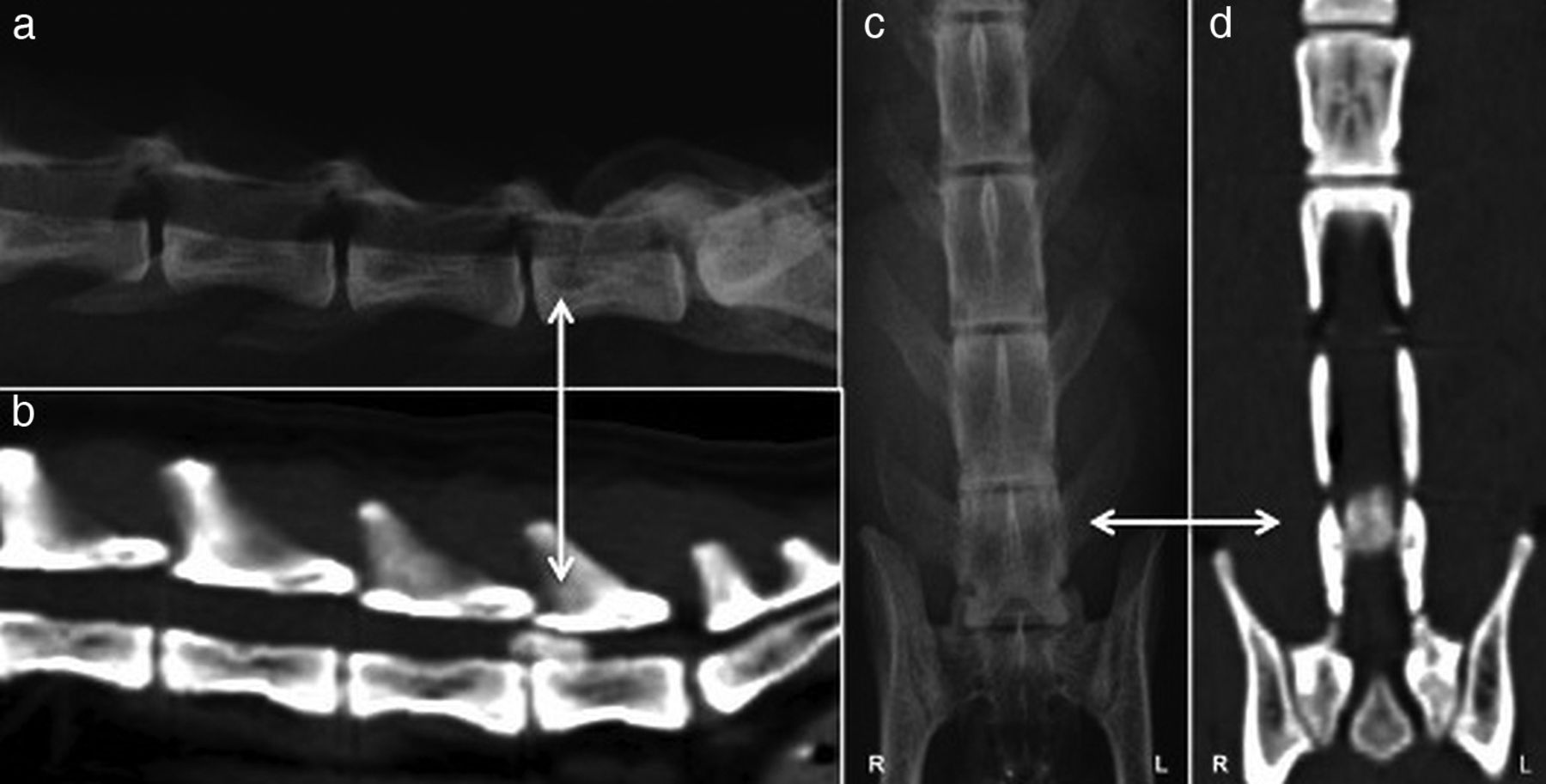
Advanced diagnostic imaging of the lumbar spine revealed caudal lumbar disc disease in both animals. MRI in the dog showed a focal bulging of the annulus fibrosus at the dorsal aspect of the L7-S1 intervertebral disc, which was causing compression of approximately 50% of the spinal canal. Both the left and right intervertebral foraminae of L7-S1 were filled with intermediate to low signal T2 weighted images (T2WI) and T1 after contrast (T1+C) material above the intervertebral disc. A CT scan of the cat revealed a large amount of hyperattentuated material within the spinal canal extending from the endplate of L6 vertebrae to the cranial third of L7 vertebra and occluding most of the vertebral canal.
The praying-position like posture and gait vanished with suitable treatment in both pets. The owner of the dog elected conservative treatment of exercise restriction and oral steroids leading to it being only mildly paraparetic. The cat underwent a L6-L7 dorsal laminectomy removing calcified intervertebral disc material resulting in resolution of nearly all neurological deficits.
It is suspected that the praying position-like posture resulted in greater pain relief for the dog and cat. Adopting a praying position-like posture results in ventroflexion of the caudal lumbar spine and decreased impingement of the nervous structures due to an increased distance between the dorsal aspects of the vertebral bodies of the adjacent vertebrae. A praying position-like posture has been previously described in different pathologies. For instance, acute abdominal pain can present with a prayer-type posture, abdominal distension, restlessness, vomiting, diarrhoea and collapse. Another report described severe cervical pain and an intermittent praying position-like posture and gait in a bulldog with C4-5 ventral cervical disc extrusion.
The two cases raise the importance of considering a praying position-like posture as clinical presentation for caudal lumbar disc disease in dogs and cats.