If you are ever faced with a juvenile hairy-nosed wombat with a limp, the recently published case report by Gail Anderson and colleagues (published in Veterinary Record Case Reports and found here) should contain a salutary lesson.
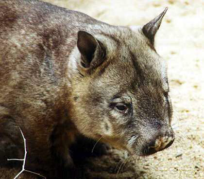
The authors were presented with a male juvenile hand-raised southern hairy-nosed wombat, which weighed 7.5 kg and was approximately 13 months old. He had been rescued from his dead mother’s pouch about seven months earlier and raised by a carer using southern hairy-nosed wombat milk replacer (yes – it does exist!). His carer had noted he was reluctant to walk and this lameness became progressively worse.
Clinical observation showed that he was reluctant to move and, when encouraged to do so, he had severe lameness in both hindlimbs and a ‘shuffling’ gait. The wombat was placed under general anaesthesia and palpation of the stifles elicited crepitus on both sides but no obvious joint effusion. It was not possible to fully extend the stifles. No other abnormalities were found on clinical examination. Stifle radiographs revealed displacement of the distal femoral metaphyses due to bilateral type 1 Salter-Harris epiphyseal fractures.
Distal femoral metaphyses have a mottled, radiolucent, appearance. Proximal femoral epiphyses were flattened and showed delayed development consistent with epiphyseal dysplasia. The right proximal femoral epiphysis was slightly more irregular and flattened compared with the left proximal femoral epiphysis. The lower lumbar spine was normal according to radiographs.
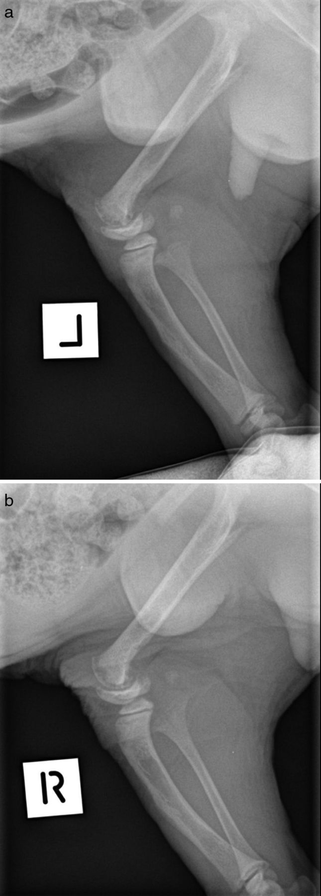
Radiographs of a normal southern hairy-nosed wombat of the same age were not available for comparison and, to the author’s knowledge, are not available in the literature. This situation is a common problem for veterinarians treating lesser-studied wildlife species.
After discussion with his carer, he was scheduled for surgery to attempt to reduce the epiphyseal fractures. If left untreated, it was unlikely that he would have regained normal mobility and function. The wombat was, however, given a guarded prognosis, partly because the injury appeared to be chronic and other radiographic changes had been observed.
Induction of anaesthesia was somewhat problematic as endotracheal intubation was difficult, probably due to the relatively small diameter of a wombat’s trachea and excessive mucus production.
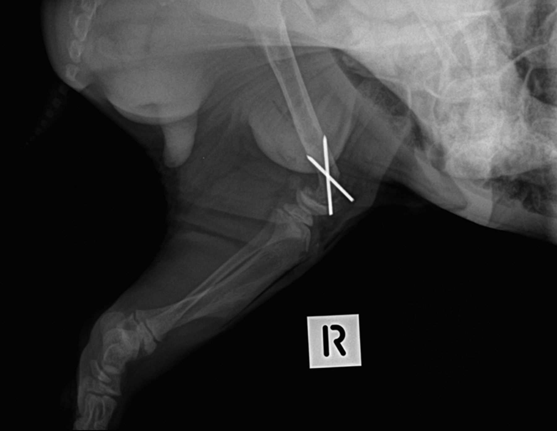
The surgical procedure was similar to that commonly used for the repair of comparable fractures in dogs. Using a small osteotome and mallet, the cartilaginous and bony epiphyseal piece was elevated and freed from its caudally displaced position and gently levered back into a position more cranially. Once reduced, the epiphyseal piece was secured with two 1.5 mm diameter Kirschner wires.
The wombat recovered quickly and uneventfully from general anaesthesia and was given postoperative analgesia. Once he was moving freely and starting to hide in its custom-made pouch (a fleece-lined pillowcase), he was left in a quiet, dimly lit cage and closely supervised.
The wombat was discharged to his carer once he was moving normally in his pouch, with instructions to restrict his activity for two weeks. He continued to eat well, although he showed initial discomfort and limited mobility. However, he continued to improve and by four months after surgery, he was walking with good extension of his hindlimbs and normal action. His carer felt that he had made a complete recovery.
A video of the wombat four months after surgery showing excellent recovery and mobility can be viewed here: www.youtube.com/watch?v=HqstN5sS8Hg.
Similar hindlimb injuries in pouch young have been frequently observed by vets working in Australia. It is thought that forcible removal of a juvenile wombat from its dead mother’s pouch is the usual cause. However, this report, adding as it does to the limited resources available for this species, shows that excellent outcomes can be achieved following this type of injury in wombats.
More details, images and discussion about this case can be found at Veterinary Record Case Reports – http://vetrecordcasereports.bmj.com/content/3/1/e000099.abstract