It is well known that the common or garden grass seed is the root of many problems in veterinary practice. Two recent articles published in Veterinary Record Case Reports (here and here) describe less common, but serious consequences following the ingestion of a grass awn by dogs.
Case 1
A 15-month-old female mixed-breed hunting dog weighing 16.5 kg was referred with a three-day history of change in bark, progressively worsening dyspnoea, decreased appetite and dullness. The onset of clinical signs was shortly after hunting. On admission, the bitch showed abduction of the forelimbs and inspiratory dyspnoea. Thoracic auscultation revealed muffled heart and dull lung sounds in the caudodorsal part of the thorax, as well as crackles on the caudal lung lobes.
Three litres of air was aspirated from the chest and supplementary oxygen was delivered via a facemask. Thoracic radiography showed elevation of the heart from the sternum and increased thoracic lucency due to the presence of pleural air. The edges of the caudal lung lobes were retracted from the chest wall and an area of soft tissue opacity was evident in the area of the caudal vena cava, the caudal border of the heart and the cupula of the diaphragm.
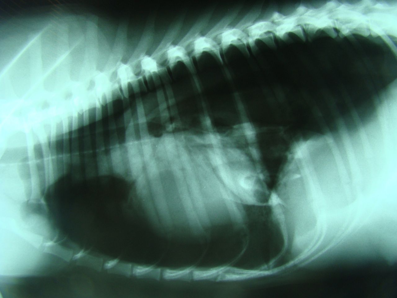
Soon after radiography the dog developed severe dyspnoea and cyanosis. Thoracentesis was repeated and 2500 cm3 of air was removed while the syringe plunger was forced back, indicating tension pneumothorax.
Tension pneumothorax is an emergency condition in which a flap of tissue acts as a one-way valve so that air entering the pleural space during inspiration cannot be expelled during expiration. This is a rapidly deteriorating state that must be recognised and treated quickly, otherwise it may prove fatal.
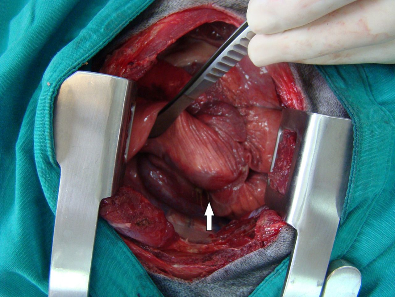
An emergency exploratory thoracotomy was performed and a grass awn was found adherent to the parietal pleura of the right 10th intercostal space and a 2 mm rupture, covered with fibrin, was identified in the ventral surface of the right caudal lung lobe. A partial lung lobectomy was performed. The dog recovered uneventfully, and was discharged two days after surgery. On re-examination two and six months later, the dog was normal and thoracic radiography revealed no abnormalities.
Case 2
In another case, a six-month-old male neutered crossbred dog presented with a two-week history of pain on opening the mouth and intermittent lethargy. There was also a history of mild blepharospasm of the right eye and pyrexia. On referral, ophthalmic examination revealed mild exophthalmia of the right eye, with a small amount of third eyelid protrusion and decreased retropulsion of the globe. There was marked pain on opening the mouth and palpation over the right temporal region of the skull.
Ocular ultrasound showed increased echogenicity of the tissue within the retrobulbar space and marked diffuse signal on Doppler examination. MRI of the brain and orbit revealed dependent fluid in the right frontal sinus and nasal cavity with mucosal thickening suggesting local rhinitis and sinusitis. There was hyperintense retrobulbar tissue thickening along the lateral aspect of the orbital lamella of the frontal bone, and evidence of regional myositis.
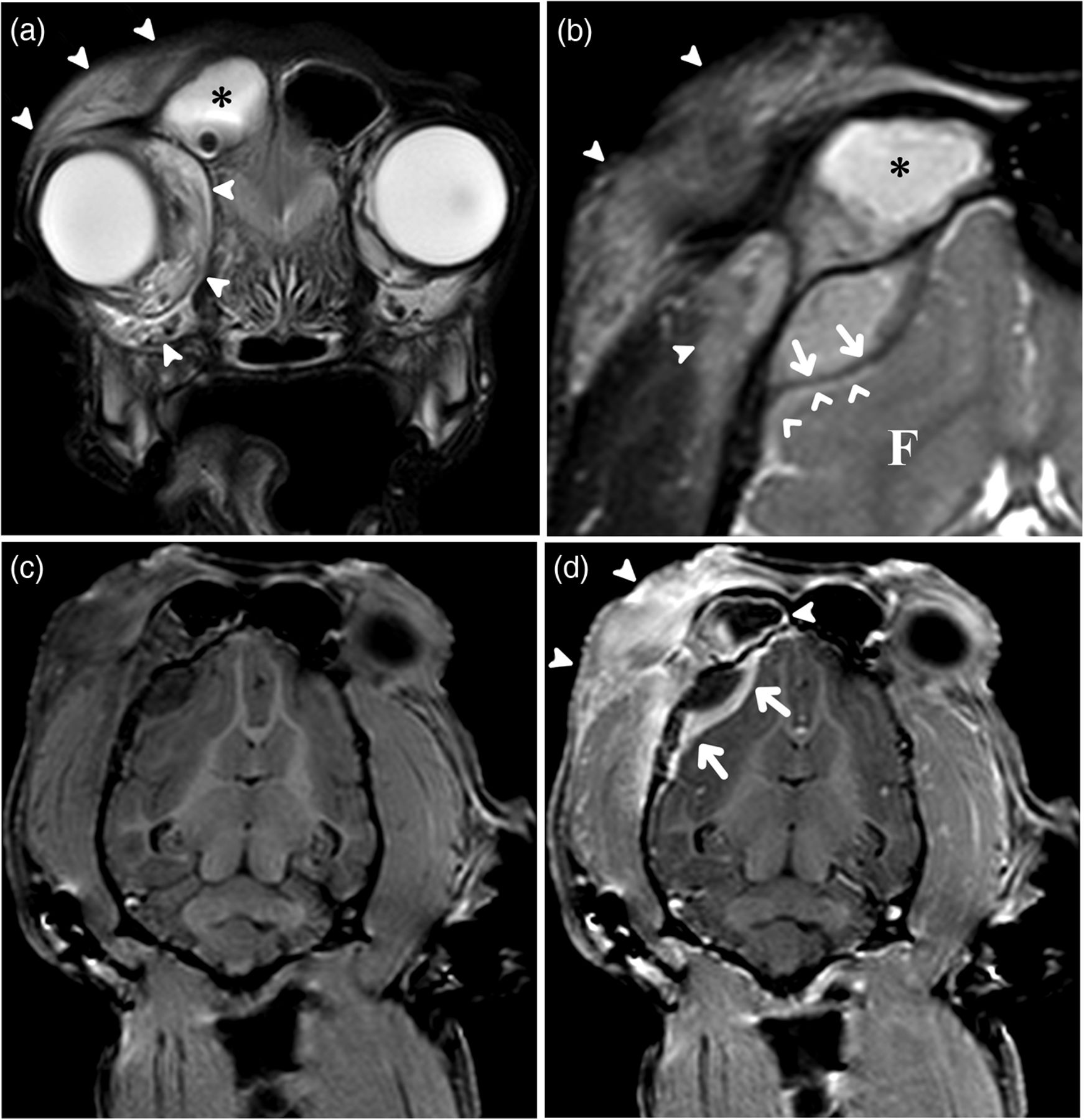
Right exophthalmos was present, likely due to diffuse retrobulbar swelling. Despite this, orbital structures were preserved.
Exploratory craniectomy was performed. And three grass seeds and approximately 4 ml of yellow, high viscosity fluid were removed from the surgical site, which was then lavaged with warm saline. Postoperatively the dog was maintained on intravenous amoxicillin and clavulanic acid before being discharged on oral medication.
Four months after the cessation of antibiotic therapy the dog was re-examined and the owner reported no problems when the dog opened its mouth. The dog was also reported to have returned to a normal life and general physical, neurological and ophthalmic examinations were unremarkable.
More details, images and discussion about these cases can be found here and here.