Veterinary Record Case Reports publishes high quality cases in all disciplines, so that clinicians and researchers can easily find important information on both common and rare conditions. Here, Alastair MacMillan, Editor of the online-only journal, highlights an interesting case involving an inquisitive labrador.
An eight-month-old female labrador retriever presented with progressive cervical hyperaesthesia after being seen coughing close to a broken sewing kit two weeks previously. She had cervical hyperaesthesia and mild proprioceptive deficits in the right thoracic and pelvic limbs. CT imaging of the neck showed a thin metallic foreign body going in a ventrodorsal direction through the vertebral canal at the atlanto-occipital junction.
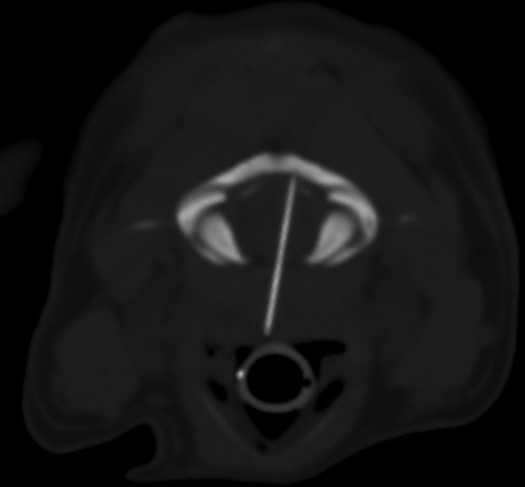
CT showing a sewing needle going in a ventrodorsal direction through the vertebral canal at the atlanto-occipital junction
Once the needle was located, it was easily grasped using Mosquito forceps, and removed in its entirety. Marked clinical improvement was observed the day after surgery and the owner reported a complete recovery of the patient, with return to normal activities in due course.
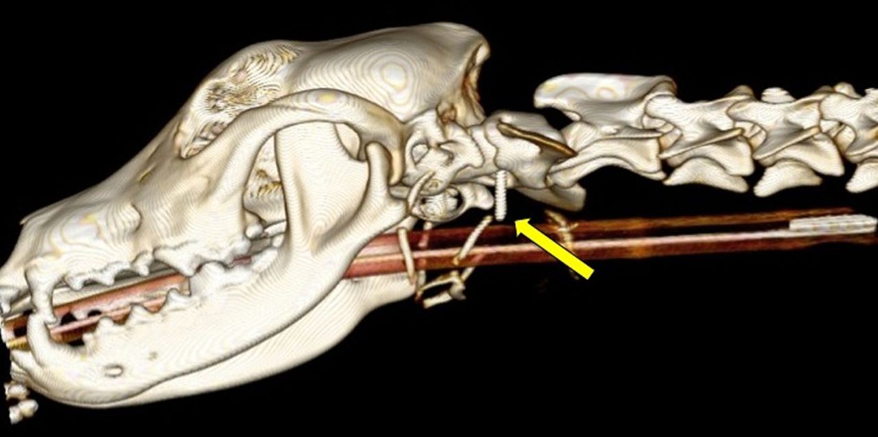 3D reconstruction showing the needle in the atlanto-occipital junction (arrow)
Reports of foreign bodies in the vertebral canal are rare in human and veterinary medicine. Although ingestion of foreign bodies is common in companion animals, sewing needles without an associated thread rarely cause a problem, as they either fail to reach the stomach, or pass through the intestinal tract uneventfully. Although brain abscessation associated with a penetrating needle has been previously reported, this is the first report of a sewing needle penetrating the vertebral canal and being surgically removed with complete clinical recovery of the patient.
To read the full report, click here.